
Hip Dysplasia
Hip Dysplasia is an inherited trait and is controlled by the
genetic make-up (genotype) of the individual dog. The genotype
is determined by the genes received from the parents - one half
from the sire and one half from the dam. The current concept is
that hip dysplasia is a polygenic trait meaning that many genes
affect that trait.
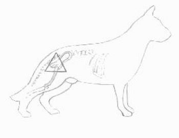
The hip joint is the part of the skeletal system that joins the
structures of the rear limb with the pelvis. It is the focal
point for transfer of driving power, generated by the rear
limbs, to the remainder of the body.
The hip is a ball and socket joint. Normal function depends on a
good fit of the ball (head of the femur) within the socket (acetabulem)
The surfaces of the head of the femur and acetabulem are covered
with smooth articular cartilage. The joint is encased by a
specialized type of connective tissue called the joint capsule
and this produces a joint fluid which lubricates and nourishes
the articular cartilage.
Normal development of the hip, from birth to maturity, is
determined by how well the parts fit together.
However, one thing is clear.
Scientists have repeatedly demonstrated that hip dysplasia is
only controllable by selective breeding and thus the need to
x-ray is indisputable.
KC/BVA SCHEME
Hip Dysplasia is neither a new disease nor one exclusive to the German Shepherd Dog as it can and does affect all breeds.
The discovery in dogs was made by an American Veterinary called Schnelle in 1935 but it attracted little attention for the next 21 years until the American Aninvale Kennels made public the fact that their dogs were affected.
The BVA/KC Hip Dysplasia scheme which had been going since 1965 had just three categories: "Certificate", "Breeder's Letter" or "Fail". Most dogs fell into the "Fail" category and as one could not tell a "near miss" from a "total disaster" it resulted in very limited use.
Around 1975 a small group of breeders got together to form the German Shepherd Dog Improvement Foundation. These were Mr & Mrs R Allan (Shootersway), Mr P Elliott (Vikkas), Mrs B. Lines (Melony), Mr H. Teall (Glenteall), and Mrs M Tidbold (Eveley). The late Dr M. Willis was the advising Geneticist for the Improvement Foundation. One of their major achievements was to establish the Hip Scoring scheme which is still in use.
A few years later the scheme was taken over by the League and was know as the GSDL/BVA Hip scoring scheme. Later still it was taken over again by the Kennel Club and remains known as it is today; the joint KC/BVA Hip Dysplasia Scheme.
Overseas countries have their own methods of determining the status of a dog's hips from an x-ray but these are not always compatible with the method used by the BVA and occasionally discrepancies occur.
The American OFA scheme started in 1966 and the German 'a' scheme in 1967.
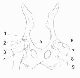
- Cranial Acetabulem Edge
- Cranial Effective Acetabulem Rim
- Dorsal Acetabular Edge
- Caudal Acetabulem Edge
- Joint Space
- Head of Femur (ball)
- Neck of Femur
- Acetabulem Fossa
- Lesser Trochanter
1-9 are the areas of the hip joint used to asses degree of dysplasia. Each hip is scored separately to give a total of 0 (lowest) to 53 (highest) thus the lowest combined total is 0 and the highest combined total is 106. The breed average today stands at a total of 19 for both hips.
Dogs cannot be x-rayed under the BVA/KC Scheme until they are 1 year old.
For further information please contact The Secretary (see Contact page).